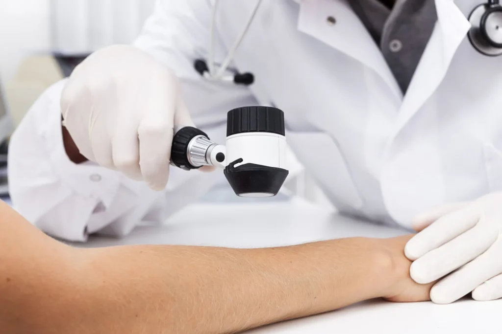
A skin cancer exam by a dermatologist is a focused, comprehensive evaluation of the skin from head to toe. This process uses clinical expertise and specialized tools to detect abnormal skin changes as early as possible. This supports skin health and allows for prompt follow-up when needed.
What Is Skin Cancer?
Skin cancer refers to the uncontrolled growth of abnormal cells in the skin’s outer layer. It typically results from DNA damage to skin cells, leading to mutations and the formation of malignant tumors. The three most common types of skin cancer each have distinct features:
- Basal Cell Carcinoma (BCC): The most common form, BCC develops from basal cells at the bottom of the epidermis. It might present as a pearl-like bump, most often on sun-exposed areas.
- Squamous Cell Carcinoma (SCC): SCC arises from squamous cells found in the outer skin layers. It typically appears as a flat lesion with a scaly surface.
- Melanoma: Though less prevalent, melanoma is the most aggressive type and arises from pigment-producing melanocytes. Signs include new, unusual growths or notable changes in an existing mole’s appearance.
What Does a Dermatologist Look For?
A dermatologist conducts a methodical skin cancer screening, examining the skin for irregularities. This careful approach increases the ability to identify features associated with skin cancer. The ABCDE method helps evaluate moles and spots for melanoma:
- Asymmetry: Unequal shape between the two halves of a spot.
- Border: Irregular, notched, or poorly defined edges.
- Color: Varied shades of tan, brown, black, or sometimes blue within a single lesion.
- Diameter: Greater than 6 millimeters, although some melanomas are smaller.
- Evolving: Change in size, shape, color, or elevation.
Dermatologists look for features associated with basal cell carcinoma and squamous cell carcinoma. The exam covers the entire body to prevent missed lesions, and can include areas like the scalp, ears, lips, palms, soles, between fingers and toes, and under the nails. This attention to less visible areas helps identify cancers that might go unnoticed.
How Is Cancer Treated?
When a suspicious lesion is detected, a dermatologist usually recommends a skin biopsy. This straightforward procedure involves removing a sample of tissue for examination by a pathologist. The microscopic analysis helps confirm whether abnormal or cancerous cells are present and guides the approach to treatment.
Treatment is tailored to the cancer’s type, size, depth, and location. For smaller or localized skin cancers, surgical excision is commonly used to remove the growth and a margin of surrounding tissue. Cryosurgery, which freezes abnormal cells, and electrosurgery, which uses electric current, are other options for some cases.
For tumors in sensitive or high-risk areas, or those that are large or have recurred, Mohs surgery may be selected. In this technique, thin layers are removed and checked microscopically until no abnormal cells remain. Mohs surgery is designed to preserve as much healthy tissue as possible. Other methods, such as photodynamic therapy, can also play a role depending on individual circumstances.
Schedule an Examination
A single, thorough skin exam by a board-certified dermatologist can help identify changes that require further monitoring or evaluation. If you detect new or changing lesions or have risk factors for skin cancer, a professional examination can support ongoing skin health and promote early intervention. They can conduct biopsies and provide treatment when it is necessary.

