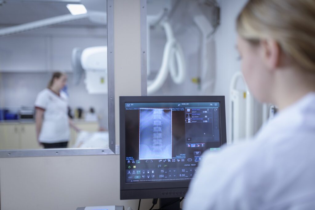
When dealing with an injury, a doctor may recommend medical imaging to get a clearer picture of what’s happening inside your body. Two common types of imaging are X-rays and magnetic resonance imaging (MRIs). While both are diagnostic tools, they serve different purposes and function differently. Understanding the differences between these two technologies can help you know what to expect from your medical evaluation.
What Is an X-ray?
An X-ray is a type of medical imaging that uses a small, safe amount of electromagnetic radiation to create pictures of the inside of your body. This radiation passes through soft tissues, such as skin and muscle, but is absorbed by denser materials, like bone. The result is a two-dimensional image that clearly shows skeletal structures.
The procedure for getting an X-ray is quick and painless. A technologist will position the part of your body being examined between the X-ray machine and a detector plate. The process only takes a few minutes, making it a fast method for diagnosing certain conditions.
What Is an MRI?
Magnetic resonance imaging (MRI) is another medical imaging technique that does not use radiation. It uses a powerful magnetic field and radio waves to generate detailed, three-dimensional images of the body’s internal structures. This includes soft tissues like muscles, ligaments, tendons, and organs.
An MRI scan takes longer than an X-ray, usually between 30 and 50 minutes. During the scan, you will lie inside a large, tube-shaped machine. The detail provided by an MRI is very high, allowing doctors to see soft tissue structures with great clarity.
How Are They Different?
The primary difference between an X-ray and an MRI lies in the technology they utilize and the types of structures they are most suited for visualizing. X-rays use ionizing radiation to produce images and are most effective for examining bones. This makes them suitable for identifying fractures and other bone-related issues.
MRIs use magnetic fields and radio waves, which means there is no exposure to radiation. This technology excels at creating detailed images of soft tissues. MRIs are preferred for examining muscles, ligaments, tendons, and internal organs.
What Are They Used For?
Doctors choose between an X-ray and an MRI based on the specific condition they need to diagnose. An X-ray is often the first step in assessing bone injuries. Common uses for X-rays include:
- Diagnosing bone fractures
- Identifying joint dislocations
- Detecting arthritis
An MRI is used when a detailed view of soft tissues is needed. It provides a more comprehensive look at structures that are not visible on an X-ray. Common uses for MRIs include:
- Diagnosing torn ligaments or tendons
- Identifying herniated discs
- Examining tumors or infections in soft tissue
How Is Professional Guidance Beneficial?
Receiving guidance from a medical professional is a key part of the diagnostic process. A specialist, such as an orthopedic doctor, can interpret the results of your imaging scan. They use their expertise to make an accurate diagnosis based on the images. This professional assessment also allows for the creation of a targeted treatment plan that can alleviate pain and promote healing.
Talk to a Specialist Today
X-rays and MRIs are both valuable diagnostic tools in orthopedic and sports medicine. X-rays offer a quick way to view bones, while MRIs provide a detailed look at soft tissues. If you have an injury or are experiencing pain, seeking professional medical advice is the next step. A specialist can guide you through the diagnostic process and help you understand your treatment options.

